
ECOCOLOURDOPPLER carotid (TSA) – Synthetic notions for the patient
WHAT
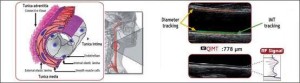
The Doppler ultrasound of the carotid or TSA is a dynamic ultrasound examination of the carotid arterial circulation / spine performed in order to assess the presence of vascular diseases borne by the district in question. It is now the gold standard (reference examination) for the diagnosis of carotid and vertebral pathologies due to the invasiveness NOT , the diagnostic accuracy and the absence of contraindications. In the past, the examination information produced solely on morphological characteristics of the vessels explored ( expiration of the arterial blood vessels , presence of aneurysm or tortuosity), on the presence of plaques ( wall thickening) their characteristics and evaluation of the degree of stenosis ( narrowing caused by plaque) as well as on the presence of glomus tumors or subclavian steal syndrome. To date, through the evolution of the technologies we can use machines that can automatically measure the thickness of the intima and media , RFQIMT ( the inner layers of the artery) and to give such elements for early diagnosis of the state of the carotid arteries with an estimate of cardiovascular risk calculated in relation to age , breed and sex. It is important that this detection is always performed on the same artery ( carotide comune) automatically ( an average of several measurements in a stretch of at least 1,5 cm), in diastole and always at the same distance from the carotid bulb. In doing so the method will be valid and reproducible. Another important parameter evaluated today only with equipment of last generation is the RFQAS ie the degree of elasticity of the artery. RFQIMT The first technology making it possible to study the thickening Intima-Media ( the inner walls of the artery) with elevata precisione. Leveraging the RF signal (Radio Frequency Data Processing) in real-time, ensures high accuracy of a few mM, thus being the method of choice for the early detection of atherosclerosis. With the technology RFQIMT is easy to determine early on the onset of diseases such as atheromatous: the intimal hyperplasia, fibrosis of the average, the early formation of small plaques, etc.. The measurements and protocol RFQIMT follow the Mannheim Consensus.RF QIMT
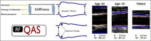
 The second technology RFQAS, always using the RF signal (Radio Frequency Data Processing), early determines the rigidity of the walls of the vessels and the method turns out to be more innovative and accurate in assessing the state of health of blood vessels. The RFQAS through a dedicated algorithm, associated parameters distension of the vessel walls to the local blood pressure, thus determining the vascular stiffness.
RF-QAS
ANATOMY OF THE WORKING cerebral arterial
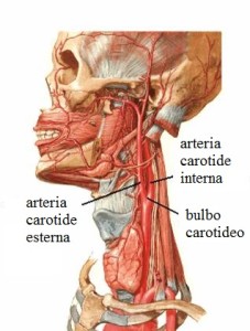 The flow of blood to the brain is guaranteed by four main arteries : The two common carotid arteries and the two vertebral. The right common carotid artery originates from the bifurcation of the trunk anonymous which in turn comes directly from the aortic arch from which it originates directly instead of the common carotid sinistra.A level of the thyroid cartilage the common carotid artery dilates forming the carotid bulb and then bifurcate in the carotid . The vertebral arteries originate from a. subclavian and after having assembled to form the basilar artery supplying the rear of the cervello.Ovviamente there may be a variety of anatomical abnormalities of course is that the source of the vessels described.
TECHNICAL D'ESAM
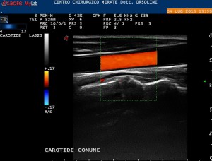
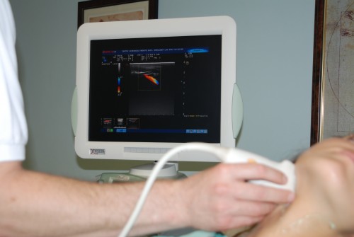
The Doppler ultrasound of the carotid is NOT an invasive examination. You run an outpatient basis and takes about 10/15 minutes. Preliminarily will be a brief history aimed at the recollection of personal data and that will be investigated mainly cardiovascular disease and previous surgical interventions , taking medication , the values of some blood tests ( total cholesterol and fractionated ) and certain dietary habits and life ( smoke). The patient is then made to lie on a table and will have to clear the neck from necklaces. Is performed at this point a detection of the blood pressure so as to interface the results with the measurement of RFQAS. Applied successively to a modest amount of conductive gel and subsequently by means of the external probes high frequency resting on the skin it will detect the ultrasonic images necessary to the diagnosis. During the examination, it is possible that the operator asks to perform movements and lateral extensions of the neck in order to better display the arterial vessels. At the end of the examination , that is completely painless , will be issued a report accompanied by representative iconography and a table with the measurements of the carotid artery elasticity (RFQAS) and the thickness of the media and intima. (RFQIMT).RESULTS
The Doppler ultrasound of the carotid is a test that should be run in the family always in the presence of cardiovascular disease such as stroke or heart attack , in smokers , obese or with high levels of total cholesterol and LDL. Obviously, any patient arteriopathic must perform the examination. In the past it was only a diagnostic method mainly because it was investigating the presence of plaques and measured the subsequent obstruction of blood. It was still very useful as a test plaque "critical" ( conditioning hemodynamically significant stenosis > 75% or irregular surface or ulcerated) needed a timely intervention by the vascular surgeon. To date with new technologies and RFQIMT RFQAS can be considered in all respects compared to a prior vascular disease may be preventable with early therapy or just with the change of lifestyle and / or food.


- Analysis of the internal carotid artery
- Carotide comune
- Flusso carotide comune
- Internal carotid artery flow
- Internal carotid
- Vertebral Artery
- Vertebral artery flow
- Flusso carotide esterna
- Carotid sternum
- Measurement QAS
- IMT measurement
Tags: carotid, Doppler ultrasound, epiaortici, examination, IMT, QAS, aortic, trunks, tsa
Trackback from your site.